
Ultra-low Radiation Dental Diagnostics
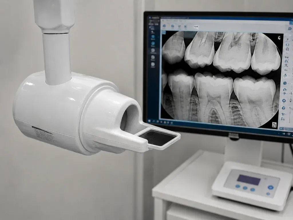
Digital radiography is a cornerstone of modern dentistry. It allows us to view anatomical structures that are completely hidden during a standard visual examination, including underneath the gums, between tight enamel contacts, and inside the jaw bones.
At Dr Batra's Dental Hub, our imaging suite is equipped with advanced digital RVG (RadioVisioGraphy) sensors and high-resolution panoramic scanners. These medical-grade devices minimize radiation exposure by up to 90% compared to old dental films, producing sharp, high-contrast digital images instantly.
Why Do We Use Digital X-rays?
- Detect Interproximal Decay: Catching early cavities developing in the tight spaces where adjacent teeth touch.
- Analyze Root Anatomy: Essential for mapping root curvatures and lengths prior to root canal treatments.
- Evaluate Bone Density: Crucial for ensuring sufficient bone height and quality before placing dental implants.
- Monitor Erupting Wisdom Teeth: Tracking the orientation and impact levels of third molars.
Why Digital Rather Than Film
Digital sensors need a fraction of the radiation of traditional film, and the image appears on the chairside screen in about two seconds rather than after processing. That means we can show you what we are looking at while you are still in the chair, and zoom or adjust contrast on the spot rather than squinting at a small film.
What X-rays Reveal That an Examination Cannot
- Decay between teeth, where the surfaces touch and no instrument can reach
- Decay developing underneath existing fillings and crowns
- The level of bone supporting each tooth — the difference between gingivitis and periodontitis
- Infection at the root tip, which indicates a tooth needs root canal treatment
- Unerupted, impacted or extra teeth, including wisdom teeth
- Cysts and other changes in the jaw that produce no symptoms
- Bone height and width for dental implant planning
Types We Use
- RVG (intraoral). A small sensor placed beside the tooth for a detailed view of one or two teeth. Used for diagnosing decay and during root canal treatment.
- OPG (panoramic). A single wide image of both jaws, all teeth, the sinuses and the jaw joints. Used for overall assessment, wisdom teeth and treatment planning.
- CBCT (3D cone beam). A three-dimensional scan showing bone volume and the position of nerves and sinuses. Used for implant planning and complex cases.
Is the Radiation Safe?
The dose from a digital dental X-ray is very small — considerably lower than film, and a fraction of the background radiation you are exposed to naturally in everyday life. We still follow the ALARA principle (As Low As Reasonably Achievable): radiographs are taken only when there is a clinical reason, never routinely just because you have attended.
If you are pregnant or think you may be, tell us before any radiograph. Non-urgent imaging is normally deferred; where an X-ray is genuinely necessary, appropriate shielding is used.
How Often Are They Needed?
There is no fixed schedule. Frequency depends on your risk of decay, gum condition and history. A patient with healthy teeth and no history of decay may need bitewings only every two to three years; someone with active decay or gum disease may need them more often. It is a clinical judgement, not a routine.
Digital X-rays in South Bopal & Ahmedabad
Radiography is carried out in-house at our South Bopal clinic, so there is no need to travel elsewhere for imaging and return with films. Images are reviewed with you on screen during the same appointment as part of your check-up. Book an appointment.